During infancy, the flexibility of the sutures, which are the points where the skull bones meet, allows the brain to grow rapidly.
However, premature closure of these bones or asymmetries caused by external factors can lead to cranial deformities.
This condition is not only an aesthetic concern but also a medical condition that carries the risk of the brain not finding space to develop.
Modern pediatric neurosurgery aims to ensure the child’s future brain development by carefully classifying these deformities.
What is Cranial Deformity in Infants? What are its Types?
Cranial deformity is the general term for abnormalities in the arrangement of the skull bones and is divided into two main categories based on its origin.
It is vitally important for parents to understand the difference between these two conditions for the accuracy of the treatment process.
Craniosynostosis: Premature Closure of the Cranial Sutures
Craniosynostosis is the premature ossification and closure of one or more sutures (growth lines) that form the skull.
In this case, the brain cannot grow in the direction of the closed suture and exerts pressure on the other open sutures, distorting the shape of the head.
This condition, which is a real surgical problem, can lead to increased intracranial pressure and restricted brain development.
Positional Plagiocephaly: Shape Deformation Due to Sleeping Position
This condition occurs when the skull flattens due to external pressure from the baby consistently lying in the same position; there is no premature closure of the bones.
It usually does not require surgical intervention; it can be corrected by changing position, using special pillows, or wearing a helmet under a doctor’s supervision.
Types of Craniosynostosis and Clinical Signs
The shape of the skull and the resulting clinical findings vary depending on which suture closes.
Scaphocephaly: Long and Narrow Skull Shape
Scaphocephaly, the most common type, occurs due to the premature closure of the sagittal suture at the center of the skull.
As the baby’s head grows forward and backward, growth to the sides stops, resulting in a “boat-shaped head” appearance.
Trigonocephaly: Triangularization of the Forehead
It is the early closure of the metopic suture in the forehead region; it is characterized by a prominent bony protrusion in the middle of the forehead and close-set eyes.
When viewed from above, the head structure resembles a distinct triangular shape.
Plagiocephaly and Brachycephaly: Asymmetries Associated with Premature Closure
Closure of the suture on one side of the head (plagiocephaly) leads to facial and ear asymmetry; closure of the posterior sutures (brachycephaly) results in a very wide and short head shape.
Diagnosis and Evaluation Process in Cranial Deformities
The diagnosis process is multifaceted to determine the need for surgery and to distinguish cases that do not require surgery.
Physical Examination and Fontanelle Check
During the specialist examination, bone protrusions (ridges) felt along the suture lines and the condition of the fontanelle are examined.
However, an open fontanelle does not always indicate craniosynostosis, as the condition of other sutures can only be determined through detailed manual examination.
Three-Dimensional (3D) Brain Tomography and Radiological Analysis
Bone-window 3D CT is used for definitive diagnosis; this method clearly shows closed sutures and the direction of brain growth.
CT images serve as a digital map for the surgeon to prepare the preoperative remodeling plan.
Craniosynostosis Surgery and Modern Approaches
Surgical intervention aims to remove the restrictive pressure on the brain and restore the skull to its most anatomically correct form.
Microsurgical Methods and Remodeling
During surgery, closed sutures are opened, and bone fragments are meticulously reshaped to create a natural head structure.
Advanced microsurgical instruments and absorbable plate-screw systems are used in this procedure to ensure that the bones fuse at the correct angle.
Timing of Surgery: What is the Optimal Age Range?
The ideal time for surgical success is generally when infants are between 3 and 9 months old; at this stage, the bones are more flexible and brain growth continues to accelerate.
In delayed cases, the operation may become more complex due to the risk of increased intracranial pressure.
Postoperative Recovery and Aesthetic Outcomes
After surgery, babies are usually monitored in intensive care for 1-2 days and in the hospital for a total of 4-5 days.
Thanks to reshaping, the baby’s head structure continues to develop naturally as they grow, and the suture marks usually fade away within the hairline.
Non-Surgical Treatment Options for Positional Abnormalities
If the problem is not premature closure of the bones, the process can be managed with non-surgical methods.
Positioning and Physical Therapy Exercises
Continuously changing the baby’s sleeping position (tummy time) distributes the pressure on the soft bones.
If the baby always looks to the same side due to stiffness in the neck muscles (torticollis), professional physical therapy support is recommended.
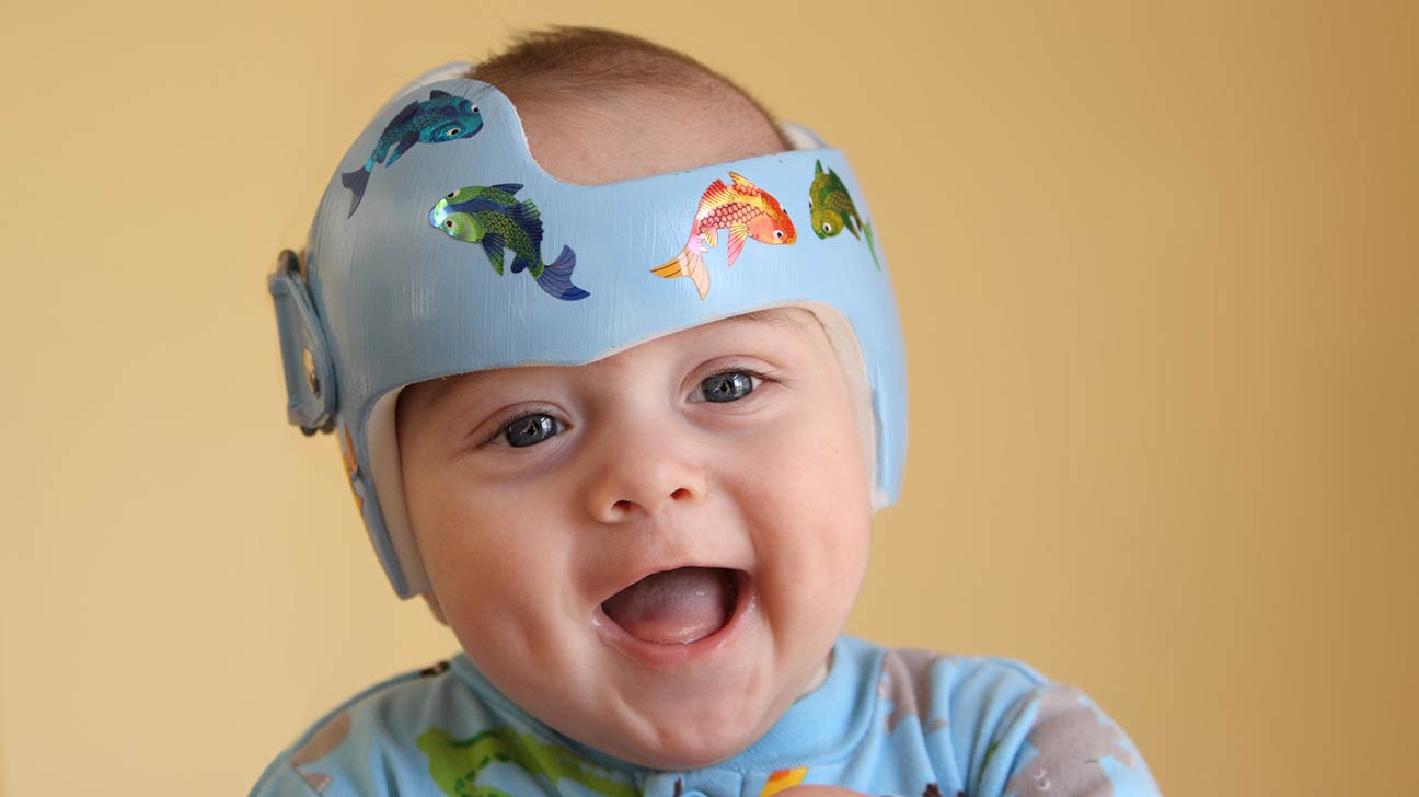
When is Helmet Therapy (Cranial Orthosis) Applied?
In cases where positional changes do not resolve the issue and significant asymmetry persists, custom-made helmets that guide the shape of the head are used.
These helmets do not apply pressure; they simply create space for the flattened parts of the head to grow, thereby guiding the bones.
Cranial Deformity Treatment and Approach Table
| Condition | Cause | Primary Treatment | Success Rate | Surgical Risk |
| Craniosynostosis | Premature closure of the bones | Surgical Remodeling | 3-9 Months | Neurosurgery |
| Positional plagiocephaly | Consistently lying in the same position | Position and Helmet | First 12 Months | None |
| Helmet Therapy | External pressure asymmetry | Cranial Orthosis | 4-12 Months | Risk of skin irritation |
| Syndromic Synostosis | Genetic factors | Multidisciplinary Surgery | Early infancy | Moderate-High |
Prof. Dr. Erdinç Özek
“The biggest mistake families make with skull deformities is to wait, thinking ‘it will correct itself over time’. If the problem is positional flattening, yes, it can correct itself; but if it is craniosynostosis, every passing month means the brain tissue remains under pressure. Clarifying the diagnosis accounts for 80% of the treatment’s success. Do not hesitate to consult a specialist pediatric neurosurgeon about even the slightest asymmetry in your baby’s head shape.”
Long-Term Effects of Skull Shape Abnormalities
Untreated cases of craniosynostosis can lead to much more profound functional losses than just an aesthetic issue.
Untreated Cases and Risks to Brain Development
When the skull does not expand, intracranial pressure increases; this can cause learning difficulties, vision loss, and developmental delays.
Compression of the brain results in the inability to use cognitive abilities to their full potential in the long term.
The Importance of Early Intervention in Terms of Aesthetic and Psychosocial Development
Cranial deformities in school-age children can lead to psychological problems such as low self-esteem and social phobia.
Anatomical correction achieved through early intervention protects both the physical and mental health of the child.
Information for Families Before and After Surgery
The greatest support for families managing this process is the transparent information provided by the surgical team.
Anesthesia Safety and Hospital Process for Infants
Today, pediatric anesthesia techniques are optimized according to the physiological characteristics of infants, minimizing risks.
Advanced technologies that prevent blood loss during surgery and the meticulous monitoring of the anesthesiologist make the process safe.
Surgical Scar and Head Circumference Monitoring
Surgical incisions are usually planned to remain within the hairline, from ear to ear; once healing is complete, the scars remain hidden under the hair.
During the first year after surgery, head circumference growth and neurological development are monitored monthly to confirm the success of the procedure.
Anonymous Case Example
A patient brought in at 4 months of age was found to have a prominent bone protrusion on the forehead and a flattening at the back of the head (trigonocephaly).
The 3D tomography results showed that the metopic suture was completely closed; the forehead area was immediately expanded using microsurgery.
At the 1-year post-operative follow-up, our patient’s head shape had returned to completely normal limits, and they demonstrated superior performance in cognitive development tests compared to their peers.
Cranial deformities are a process that can be resolved with the right steps taken at the right time.
It is essential not to compromise on scientific approaches for your baby’s healthy development and aesthetic future.
For a more detailed evaluation on the subject, you can consult an expert opinion and schedule an appointment at our clinic.
Scientific References
PubMed / NCBI: “Positional plagiocephaly vs craniosynostosis: A diagnostic challenge”. https://pubmed.ncbi.nlm.nih.gov/28365412/
Child’s Nervous System: “Surgical techniques in the management of sagittal synostosis”. https://link.springer.com/article/10.1007/s00381-020-04561-2
Lancet Child & Adolescent Health: “Early detection and intervention in pediatric skull deformities”.