Hydrocephalus is a condition in which cerebrospinal fluid (CSF) accumulates excessively in the brain ventricles due to a malfunction in the production, circulation, or absorption stages.
This accumulation leads to increased intracranial pressure, creating serious pressure on brain tissue and directly affecting neurological functions.
What is Hydrocephalus? (Fluid Buildup in the Brain)
The brain and spinal cord float in a clear fluid (CSF) that is constantly renewed and circulates.
This fluid protects the brain from impact, transports nutrients, and cleanses waste products.
However, a blockage or absorption disorder in this system causes the fluid to become trapped in cavities called ventricles and leads to enlargement of the brain.
Cerebrospinal Fluid (CSF) Cycle and Function
CSF is produced in areas of the brain called the choroid plexus and reaches the brain surface through channels between the ventricles.
Here, it is reabsorbed into the bloodstream via arachnoid villi.
When the balance of this fluid, approximately 500 ml of which is produced daily, is disrupted, hydrocephalus develops.
What Causes Hydrocephalus?
Three main mechanisms play a role in the development of hydrocephalus:
Obstruction: Narrowing or mass pressure in the fluid pathways.
Absorption Disorder: Closure of the channels where the fluid should be absorbed due to infection or bleeding.
Excessive Production: Rarely, the fluid is secreted in much greater quantities than normal.
What Are the Symptoms of Hydrocephalus?
Symptoms vary greatly depending on the flexibility of the skull and the age of the patient.
Symptoms Seen in Infants and Children
Since the skull bones have not yet fused in infants, the most obvious sign is rapid growth of the head circumference.
Noticeable tension or protrusion in the fontanel (soft spot).
Constant downward gaze of the eyes (sunset sign).
Feeding difficulties, projectile vomiting, and excessive irritability.
Symptoms Observed in Adults and the Elderly
In adults, since the skull bones are fused, increased pressure causes severe headaches and visual disturbances.
Severe headache and nausea that worsen upon waking.
Double vision or blurred vision (sign of papilledema).
Impaired balance and sudden urinary incontinence.
Characteristics of Normal Pressure Hydrocephalus (NPH)
It is commonly seen in individuals over the age of 60 and is often confused with Alzheimer’s or Parkinson’s disease.
It manifests itself with the characteristic triad of “gait disturbance, urinary incontinence, and mild dementia (forgetfulness)”.
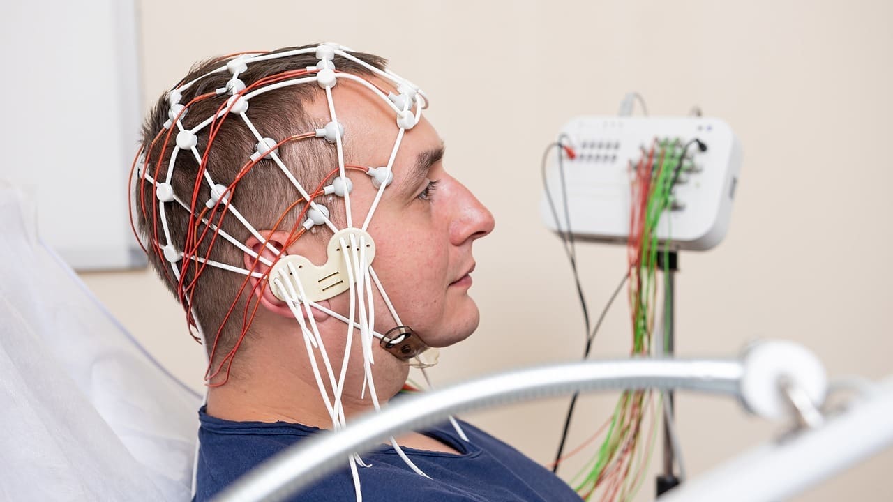
Hydrocephalus Diagnosis and Diagnostic Methods
Modern diagnostic techniques not only confirm the presence of the disease but also determine where the fluid is blocked for surgical planning.
Brain MRI and Computed Tomography (CT) Scans
Magnetic Resonance Imaging (MRI) is the gold standard for measuring the size of the ventricles and the flow rate of the fluid.
In particular, CSF flow MRI imaging provides a functional map of the obstruction, enabling a decision on whether to use a shunt or an endoscopic method.
Lumbar Puncture (Fluid Drainage Test) and Important Data
Especially in cases of suspected Normal Pressure Hydrocephalus, a small amount of fluid is extracted from the lumbar region to observe improvement in the patient’s symptoms.
This test serves as a simulation that shows in advance how effective shunt surgery will be for the patient.
Hydrocephalus Treatment Options
Hydrocephalus treatment is primarily surgical; however, the patient’s clinical condition determines the choice of treatment.
Treatment Method Comparison Table
| Feature | Shunt Surgery (VP Shunt) | Endoscopic Method (ETV) |
| Technique | A permanent tube/pump system is implanted. | A new fluid pathway is created within the brain. |
| Foreign Body in the Body | Yes, it remains in place for life. | No, it does not contain a foreign object. |
| Success Factor | It is effective in almost all cases. | It is more successful in cases related to blockage. |
| Follow-up | Regular shunt adjustment and monitoring are required. | Requires less surgical follow-up. |
Recommendations from Prof. Dr. Erdinç Özek
“Shunt systems used in the treatment of hydrocephalus are now equipped with externally adjustable (programmable) valves thanks to advances in technology.
This allows us to adjust the shunt’s pressure settings in a clinical setting without surgery as the patient’s intracranial pressure changes.
Especially in our pediatric patients, these adjustable systems are vital for meeting pressure needs related to growth.
Remember that the shunt is not an obstacle, but a supporter of a healthy life.”
What is a Shunt Surgery (Ventriculoperitoneal Shunt)?
Shunt surgery is a drainage system that removes accumulated fluid from the brain and drains it to another part of the body (usually the abdominal cavity).
How Does the Shunt System Work? (Pump and Catheter Mechanism)
The system consists of three main parts: a catheter that collects fluid from the brain, a valve (pump) that determines the direction and amount of fluid flow, and a thin tube that carries the fluid to the abdomen.
The valve mechanism opens only when the pressure rises above a certain level, preventing excessive drainage from the brain.
How is Shunt Surgery Performed? (Surgical Technique)
The approximately 1-hour procedure is performed under general anesthesia.
The system is completely placed under the skin through small incisions made in the scalp and abdomen; when viewed from the outside, it is only noticeable as a slight bulge.
Alternative Method: Endoscopic Third Ventriculostomy (ETV)
In some cases of obstruction-based hydrocephalus, it is possible to resolve the issue without placing a foreign object in the body.
Who is Shuntless Hydrocephalus Surgery Suitable For?
This method is particularly preferred in patients with interventricular canal stenosis (aqueductal stenosis).
Using a camera called an endoscope, the obstruction in the brain’s fluid is bypassed, and a new pathway is created.
Clinical Experiences and Anonymous Case Example
Case Analysis: A 72-year-old male patient presented with complaints of difficulty walking and confusion.
His family thought he had Alzheimer’s disease, but a brain MRI revealed enlarged ventricles but normal brain pressure (NBH).
A lumbar puncture (LP) showed a significant improvement in the patient’s walking ability.
Recovery and Follow-up After Shunt Surgery
The postoperative process focuses on the shunt’s adaptation and managing the risk of infection.
First 24 Hours: The patient is kept under neurological observation, and the drainage rate of the shunt is monitored.
Hospital Stay: Discharge procedures are usually completed within 2-3 days.
Signs of Shunt Dysfunction: If the patient experiences severe headache, numbness, or redness at the suture sites, this may indicate that the shunt is blocked or infected.
Shunt Revision: If the shunt malfunctions mechanically or becomes blocked, revision surgery may be planned to replace the damaged part of the system.
Hydrocephalus Frequently Asked Questions
Does shunt surgery damage the brain?
Shunt surgery is not performed to damage brain tissue; on the contrary, it is performed to eliminate the destructive pressure of accumulated fluid on the brain and prevent permanent damage.
Can a person with a shunt engage in sports?
Patients with shunts can generally engage in light to moderate sports, but should avoid heavy contact sports that could damage the mechanical structure of the shunt or pose a risk of impact.
What is the lifespan of a shunt, and when is it replaced?
Shunts are technically designed to last a lifetime, but they do not need to be replaced unless a problem such as blockage, infection, or mechanical failure develops.
What is a programmable shunt, and why is it preferred?
Programmable shunts are systems that allow pressure adjustments to be made externally using a magnetic device without the need for surgery, enabling treatment optimization according to the patient’s changing needs.
Is endoscopic third ventriculostomy (ETV) suitable for everyone?
The ETV method is only successful in patients with physical blockages (obstructions) in the fluid pathways within the brain; a shunt is usually necessary in cases of absorption disorders.
Do airport security gates or MRI machines damage the shunt?
Most modern adjustable shunts are MRI-compatible, but some models may require re-adjustment of pressure settings after an MRI; airport detectors generally do not affect shunt settings.
Hydrocephalus and Shunt Surgery Specialist Appointment
You can request an appointment at our clinic for a professional approach to hydrocephalus diagnosis, symptom management, or shunt checks.
Based on your current imaging results and clinical examination findings, Prof. Dr. Erdinç Özek carefully evaluates the latest adjustable shunt technologies or endoscopic treatment options.
You can consult with a specialist to answer your questions and create a permanent treatment plan.
Scientific References
The Lancet Neurology: Advances in the management of hydrocephalus. https://www.thelancet.com
Journal of Neurosurgery (JNS): Ventriculoperitoneal shunt versus endoscopic ventriculostomy. https://thejns.org
PubMed (NIH): Pathophysiology of normal pressure hydrocephalus. https://pubmed.ncbi.nlm.nih.gov