Head and spinal injuries are critical injuries that directly affect the central nervous system and can leave permanent effects on vital functions.
Thanks to the technological capabilities offered by modern medicine and microsurgical techniques, success rates in post-trauma interventions are increasing day by day.
In brain surgery practice, these cases are considered emergencies that require a multidisciplinary approach, where every second counts.
Accurate diagnosis, rapid surgical intervention, and a meticulous rehabilitation process form an indispensable whole for preserving patients’ quality of life.
What is Head Trauma? Classification by Severity
Head trauma is damage to brain tissue, blood vessels, or skull bones resulting from physical force to the skull.
The effects of trauma can range from a simple swelling to damage extending deep into the brain tissue.
Mild Head Trauma and Concussion
These are injuries that typically do not involve loss of consciousness or involve only very brief (a few minutes) confusion.
Patients may experience temporary confusion, dizziness, or short-term memory problems; however, neurological examinations are usually normal.
Clinical Presentation in Moderate and Severe Head Injuries
These injuries are characterized by prolonged loss of consciousness, severe projectile vomiting, seizures, and weakness in part of the body.
In these cases, the risk of cerebral edema or intracranial hemorrhage is very high; therefore, the option of surgical intervention should be kept on the table, and the patient should be monitored in intensive care conditions.
Traumatic Brain Injuries and Surgical Intervention
Since brain tissue is located inside the skull, which is a closed box, any bleeding or edema causes compression of the brain.
Epidural and Subdural Hematomas (Brain Bleeds)
Epidural hematomas are usually rapid arterial bleeds accompanying skull fractures, while subdural hematomas are caused by the rupture of venous vessels on the surface of the brain.
In both cases, the removal of the blood clot (hematoma) exerting pressure on the brain tissue through microsurgery is the key factor directly determining the chances of survival.
Treatment of Skull Fractures and Depressed Fractures
If a bone fragment has depressed into the brain tissue (depressed fracture), this can lead to tissue damage and infection.
Surgical removal of bone fragments, cleaning, and restoration with titanium plates if necessary are planned.
Intracranial Pressure (ICP) Management and Decompressive Craniectomy
In cases of severe brain edema that does not respond to drug treatment, temporary removal of part of the skull (decompressive craniectomy) may be necessary.
This procedure allows the brain to expand outward, preventing the crushing of vital centers.
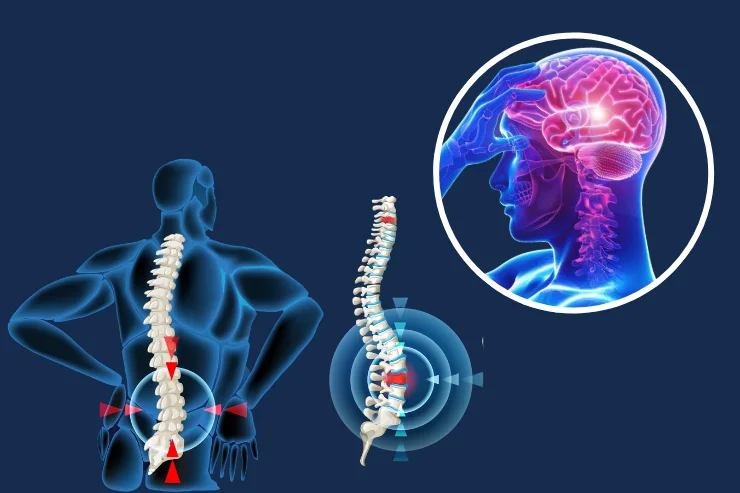
Spinal Cord and Spinal Column Injuries
Spinal injuries directly threaten the spinal cord, which runs through the spine and controls the body’s motor functions.
Spinal Fractures and Dislocations: Symptoms and First Aid
Severe back/lumbar pain, numbness, and tingling following a fall from a height or a traffic accident are early signs of a spinal fracture.
A patient who is moved incorrectly may face the risk of permanent paralysis due to an unstable fracture.
Approaches to Spinal Cord Injuries and the Risk of Paralysis
Contusions (bruises) or complete tears in the spinal cord lead to loss of sensation and movement.
Early surgical decompression (removal of pressure) aims to maximize the potential for nerve recovery.
Comparison Table for Head and Spinal Cord Injuries
| Type of Trauma | Critical Symptom | Primary Diagnostic Tool | Emergency Intervention | Recovery Potential |
| Epidural Hemorrhage | Fluctuating Consciousness | Non-contrast Brain CT | Emergency Hematoma Drainage | High in early intervention |
| Spinal Fracture | Severe pain, loss of strength | CT and MRI | Stabilization / Plating | Depends on the level of injury |
| Concussion | Dizziness, forgetfulness | Clinical Observation | Rest and Follow-up | Usually complete recovery |
| Collapse Fracture | Depression in the skull | Direct X-ray / CT | Elevation (Lifting) | Depending on the condition of the tissue |
Diagnosis and Imaging Methods in Traumas
Fast and accurate imaging is the most critical step in guiding the surgeon’s approach.
Emergency Department Management: The Importance of CT and MRI Imaging
Computed Tomography (CT) is the first choice in emergency departments because it provides results within seconds for bone structures and acute bleeding.
Magnetic Resonance Imaging (MRI) is used after stabilization to detect microscopic damage in brain tissue and the spinal cord.
Neurological Assessment and the Glasgow Coma Scale (GCS)
The GCS scoring, which assesses the patient’s level of consciousness, eye movements, and motor responses, standardizes the severity of the trauma.
A decrease in the score indicates that the surgical team should switch to a more aggressive treatment protocol.
Surgical Treatment and Stabilization in Spinal Injuries
When the injured spine can no longer bear the body’s weight, surgical stabilization becomes necessary.
Spinal Instrumentation and Plate Applications
Screw and rod (plate) systems are used to stabilize fractured or displaced vertebrae.
This procedure not only relieves pressure on the nerves but also allows the patient to get up and walk in the early stages.
Minimally Invasive Spinal Surgery and Kyphoplasty
In suitable cases, fractures can be stabilized by injecting bone cement (kyphoplasty) using closed methods without making large incisions.
This method significantly shortens the recovery time, especially in elderly patients or individuals with a high-risk general health condition.
Prof. Dr. Erdinç Özek: “The golden rule to remember when dealing with head and spinal trauma is not to move the patient. Unprofessional assistance given to a patient with suspected spinal injury can cause lifelong paralysis. For us surgeons, success begins not in the operating room, but with proper first aid at the scene.”
Post-Trauma Recovery and Rehabilitation Process
Surgical intervention is only the first step in the process; recovery of brain and nerve tissue takes time.
Intensive Care Monitoring and Vital Sign Tracking
Controlling intracranial pressure and balancing body temperature in the postoperative period are critical to preventing secondary brain damage.
Adequate oxygenation and blood pressure control ensure the nourishment of healing tissues.
Neurorehabilitation: The Role of Physical Therapy in the Early Stages
The earlier physical therapy begins in cases of nervous system trauma, the faster the brain builds new pathways (plasticity).
Preventing muscle atrophy and maintaining joint mobility are key to long-term success.
Anonymous Case Example
A 45-year-old patient brought to the emergency room after a fall from a height was diagnosed with a fractured lumbar vertebra compressing the spinal canal.
The patient underwent microsurgical decompression and posterior stabilization (plate placement) surgery within the first 6 hours after the accident.
Our patient, who had severe weakness in his legs before surgery, was able to stand with support on the third day after surgery and regained independent walking ability at the end of the sixth month with an intensive physical therapy program.
Head and spinal injuries require expertise and composure.
Regardless of the type of trauma, a treatment program planned based on scientific data can open the door to miracles.
For detailed information about the process and professional advice, you can seek support from our team of experts and schedule an appointment at our clinic.
Scientific References
The Lancet Neurology: “Traumatic brain injury: integrated approaches to improve prevention, clinical care, and research”. https://www.thelancet.com/journals/laneur/article/PIIS1474-4422(17)30371-X/fulltext
Journal of Neurosurgery: Spine: “Management of acute traumatic spinal cord injury”. https://thejns.org/spine/view/journals/j-neurosurg-spine/21/1/article-p42.xml
PubMed / NCBI: “Pathophysiology of secondary brain injury and therapeutic perspectives”. https://pubmed.ncbi.nlm.nih.gov/31256372/
World Neurosurgery: “Surgical vs Non-Surgical Treatment of Thoracolumbar Burst Fractures”.
New England Journal of Medicine (NEJM): “Decompressive Craniectomy in Diffuse Traumatic Brain Injury”. https://www.nejm.org/doi/full/10.1056/NEJMoa1102077