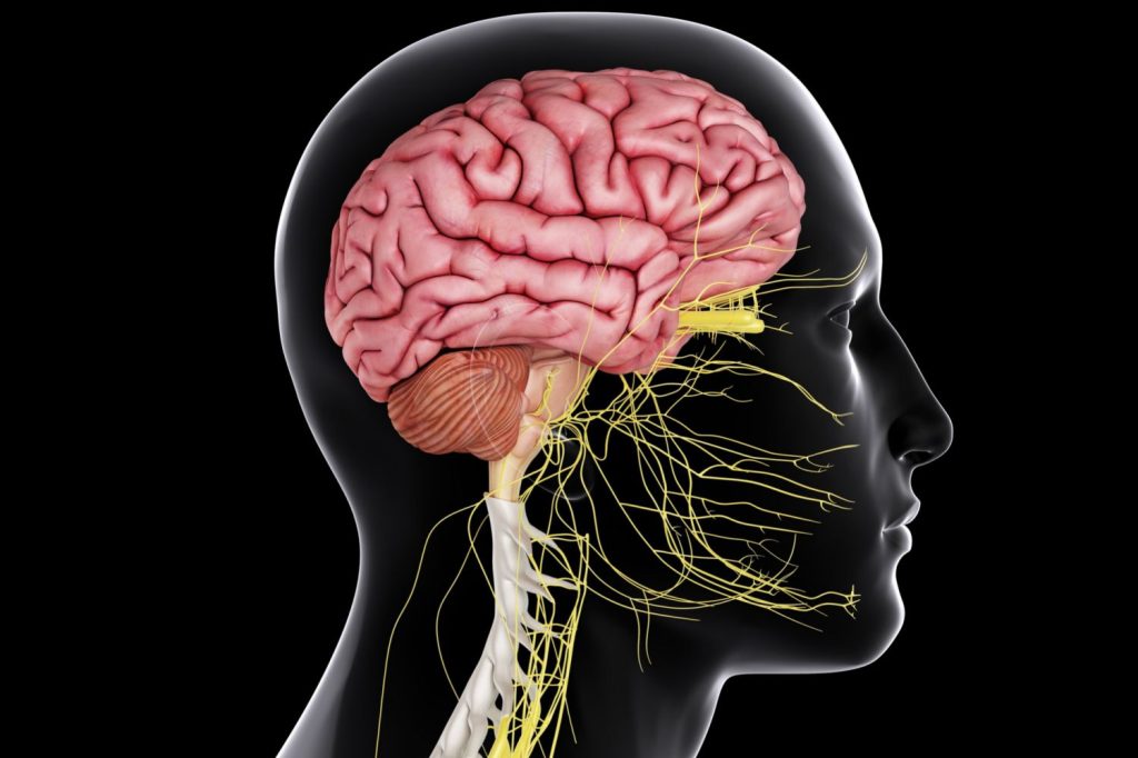
Pediatric brain and spinal cord disorders are a highly sensitive field of medicine that encompasses the entire process from the womb to the end of adolescence.
Unlike adult brain surgery, pediatric neurosurgery requires working on a constantly growing and developing nervous system.
Children’s physiological responses are much faster and more variable than those of adults; this situation requires a special approach at every stage of surgical planning.
Technological advances in modern medicine have made it possible to successfully treat many congenital or acquired anomalies that were once considered risky.
What is Pediatric Neurosurgery? How is it Different from Adult Surgery?
Pediatric neurosurgery is a specialty that deals with brain, spinal cord, and nervous system surgery in individuals from newborns to 18 years of age.
Since children’s nervous systems are still developing, surgical intervention should not only solve the current problem but also protect the child’s future development.
A Delicate Approach and Microsurgery in Pediatric Brain Surgery
In children, tissues are much smaller, more delicate, and more sensitive to blood loss.
Therefore, in pediatric cases, micro-surgical techniques and special magnifying microscopes are used to minimize the margin of error.
Everything from the anesthesia techniques used during surgery to the size of the surgical instruments is selected to suit the child’s anatomy.
The Vital Importance of Early Diagnosis in the Developmental Process
Early intervention during the first years of life, when brain development is most rapid, can preserve a child’s motor and cognitive skills.
Especially in cases where intracranial pressure increases or neurological development is compromised, delay can lead to irreversible losses.
Craniosynostosis in Children
Infants have flexible joints called “sutures” between their skull bones; these structures allow the brain to grow.
Craniosynostosis is the premature ossification and closure of one or more of these joints.
Premature Closure of the Fontanelle and Fusion of the Skull Bones
Early closure of the fontanelle and sutures prevents the skull from growing normally in all directions and causes noticeable deformities in the head (triangular head, long head, etc.).
This condition is not only an aesthetic problem, but also means a reduction in the space required for the brain to expand.
Deformity and Pressure on Brain Development
Depending on the location of the closed suture, brain tissue may be under pressure; this can lead to increased intracranial pressure, visual impairments, and developmental delays.
In cases diagnosed early, surgical outcomes are quite encouraging.
Endoscopic and Open Craniosynostosis Surgery
In infants between 3-6 months of age, only the closed suture can be opened using endoscopic (closed) methods.
In older infants, open surgical techniques are used to reshape the skull, creating space for the brain to grow comfortably.
Hydrocephalus (Accumulation of Fluid in the Brain) in Childhood
Hydrocephalus is the enlargement of the brain cavities resulting from an imbalance in the cerebrospinal fluid (CSF) circulating within the brain and spinal cord.
Symptoms of Hydrocephalus: Head Circumference Growth and Vomiting
Since the skull bones in infants have not yet fused, the most typical symptom is rapid enlargement of the head circumference.
In addition, swelling of the fontanelle, vomiting, constant drowsiness, and downward slanting of the eyes, known as “sunset sign,” may be observed.
Shunt Surgery and Modern Shunt Technologies
A shunt system is a thin tube and valve mechanism that directs excess fluid from the brain to the abdominal cavity or around the heart.
Thanks to adjustable valve technologies today, the fluid flow rate can be optimized according to the patient’s needs through external intervention after surgery.
ETV (Endoscopic Third Ventriculostomy): Shunt-Free Treatment Method
In suitable cases, treatment can be provided without implanting a foreign object (shunt) into the body by creating an endoscopic opening that bypasses the blockage within the brain.
Pediatric Neurosurgery Treatment Approaches Comparison Table
| Disease Type | Primary Symptom | Primary Treatment Method | Recovery Process |
| Hydrocephalus | Head enlargement, vomiting | Shunt or Endoscopic VENT | 1-2 weeks, lifelong follow-up |
| Craniosynostosis | Abnormal head shape | Cranial reshaping | 2-4 weeks |
| Spina Bifida | Pouch or opening in the back | Microsurgical Repair | Intensive post-operative follow-up |
| Epilepsy Surgery | Refractory Seizures | Focal resection or VNS | Variable, long-term follow-up |
Spinal Defects and Congenital Anomalies: Spina Bifida
Commonly known as “open spine,” spina bifida occurs when the spinal cord fails to close properly during the first month of pregnancy.
Meningocele and Meningomyelocele Treatment Processes
Meningomyelocele, a condition in which the spinal cord is outside the body in a sac, must be surgically closed within the first 24-48 hours after birth.
The purpose of this intervention is to protect the nerve tissue from infection and preserve existing functions.
Tethered Cord Syndrome Symptoms and Surgery
This condition occurs when the lower end of the spinal cord becomes tethered to the spinal canal during growth.
It may present with discoloration of the back, increased hair growth, or deformity of the feet; treatment involves surgical release of the tension.
Lipomeningocele: Approach to Fat Masses in the Spinal Cord
These are fatty masses located in the spinal cord and intertwined with nerves.
The goal is to reduce these masses and relieve pressure on the nerves using delicate microsurgical techniques without disrupting nerve transmission.
Prof. Dr. Erdinç Özek: “Brain and spinal cord surgery in children is not just an operation, it is a process of building a future. Babies’ nervous systems have tremendous capacity for regeneration. Our job is to remove the obstacles to that development with microsurgical precision. When touching a child’s life, the greatest power in the surgeon’s hands is the combination of scientific rigor and compassion.”
Childhood Brain Tumors and Surgical Treatment
Brain tumors in children have different locations and biological characteristics compared to those in adults.
Diagnosis and Microsurgical Planning in Pediatric Brain Tumors
Most tumors are located in areas close to the balance center, known as the posterior fossa.
Using neuronavigation and neuromonitoring technologies, the priority is to avoid damaging healthy brain tissue while removing the tumor.
Posterior Fossa Tumors and Approaches That Preserve Quality of Life
Preserving the child’s motor skills and speech abilities after surgery is as important as completely removing the tumor.
A long-term treatment plan is developed with the support of a multidisciplinary team (oncology, radiotherapy, neurosurgery).
Surgery for Drug-Resistant Childhood Epilepsy
Surgical options are evaluated for seizures that do not respond to medication and originate from a specific focus in the brain.
Identifying the Epilepsy Focus and Functional Surgery
Advanced MRI techniques and EEG mapping are used to identify the point of origin of the seizure.
If this point is not in a vital area such as the speech or movement center, removing the focus may stop the seizures.
Vagus Nerve Stimulator (VNS) and Brain Pacemaker Applications
In cases where the focus cannot be removed, a pacemaker implanted in the vagus nerve in the neck can reduce seizure frequency by sending signals to the brain.
Arachnoid Cysts and Cerebrospinal Fluid Cysts
Arachnoid cysts, which are fluid-filled sacs that form between the brain membranes, do not always require surgical intervention.
Should Cysts Be Monitored or Treated Surgical Intervention?
Many cysts are discovered incidentally and are only monitored as long as they do not grow.
However, cysts that exert pressure on the brain, trigger seizures, or increase intracranial pressure require intervention.
Cyst Treatment with Fenestration and Shunting Methods
Draining the cyst contents into the brain cavities (fenestration) or draining the cyst using a shunt are among the modern treatment methods.
Anonymous Case Example
A 2-month-old infant was brought to our clinic due to noticeable triangulation of the head and a protrusion on the forehead (trigonocephaly).
Examinations revealed that the frontal suture had closed prematurely.
Using a minimally invasive (small incision) technique, the suture was opened to provide the brain with space to grow.
At the 6-month follow-up, it was observed that the head shape had completely returned to normal and the infant’s neurological development was consistent with that of peers.
Neurological problems in childhood can be a challenging process that affects the entire family.
With the correct diagnosis and modern surgical interventions performed by specialists, children can take steps toward a healthy future.
If you have any concerns about your baby’s or child’s development, you can consult a specialist pediatric neurosurgeon without delay.
You can schedule an appointment at our clinic to initiate a detailed evaluation process.
Frequently Asked Questions
Does premature closure of the fontanelle cause intellectual disability?
Premature closure of the fontanelle or cranial sutures, if left untreated, can put pressure on the brain and negatively affect cognitive development and vision; however, early surgical intervention can prevent these risks and ensure normal development.
Do shunts have to remain in place for life in infants?
Shunt systems used in the treatment of hydrocephalus generally remain in the patient’s body for life, continuing to drain cerebrospinal fluid; however, in rare cases, the shunt may no longer be needed with advancing age, or there may be a chance to remove the shunt using methods such as ETV.
Is brain surgery risky for children?
Thanks to modern microsurgical techniques, neuromonitoring, and advanced anesthesia techniques, the risks associated with pediatric brain surgery are much lower than in the past; the harm caused to the child’s development by delayed treatment is much greater than the risk of the operation itself.
Does the head shape completely return to normal after surgery?
After craniosynostosis surgery, the shape of the skull largely returns to normal; especially in interventions performed within the first year, when bone growth is rapid, the natural pressure exerted by the brain from within helps the skull to improve aesthetically.
Can a child born with spina bifida walk?
The potential to walk depends on the level of the opening in the spinal cord and the degree of nerve involvement; with successful surgical repair in the early stages and subsequent regular physical therapy, many children can gain the ability to walk.
Can an arachnoid cyst in the brain rupture?
Arachnoid cysts generally have a strong membrane structure and do not rupture spontaneously; however, in rare cases, severe blows to the head can cause intracystic bleeding or rupture of the cyst. Therefore, children with cysts are advised to avoid contact sports.
Scientific References
The Lancet Child & Adolescent Health: “Global burden of pediatric brain and spinal cord tumors”. https://www.thelancet.com/journals/lanchi/article/PIIS2352-4642(19)30001-6/fulltext
Journal of Neurosurgery: Pediatrics: “Long-term outcomes of tethered cord release in children”. https://thejns.org/pediatrics/view/journals/j-neurosurg-peds/25/1/article-p54.xml
PubMed / NCBI: “Management of Arachnoid Cysts in the Pediatric Population”. https://pubmed.ncbi.nlm.nih.gov/30256247/