Endoscopic skull base surgery, one of the most advanced disciplines in modern neurosurgery, allows access to complex lesions in the base of the brain through natural pathways (nostrils) without opening the skull.
What is Endoscopic Skull Base Surgery?
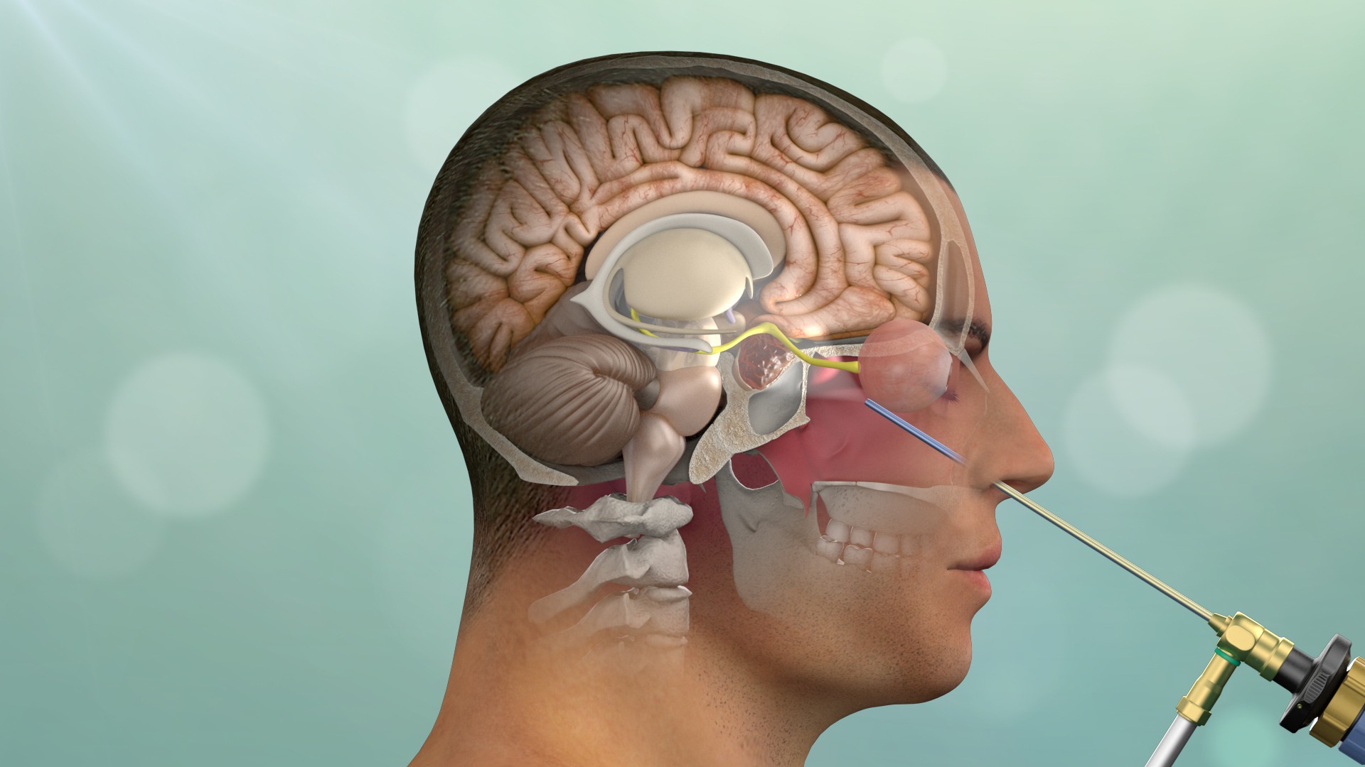
This method is based on the principle of reaching the skull base by entering through the nasal cavity, known as the “transnasal transsphenoidal” approach. Unlike classic open surgeries, there is no need to move brain tissue aside.
Minimally Invasive Approach and Technological Infrastructure
High-resolution 4K endoscopes provide the surgeon with a wide field of view. Neuronavigation devices used during surgery help the surgeon advance with millimeter precision in the depths of the brain, minimizing the margin of error.
Diseases Treated with Endoscopic Methods
Skull base surgery covers a wide range of pathologies due to the complex anatomy of the skull base.
Pituitary Adenomas: One of the most commonly treated areas; it allows for the removal of masses that cause hormonal imbalances or vision loss.
Craniopharyngiomas: Complex cystic tumors, particularly seen in children and young adults.
CSF Fistula Repair: Repair of cerebrospinal fluid leakage from the nose following head trauma or surgery.
Meningioma and Chordoma: Masses originating from the bone and membrane structures of the skull base.
Comparison of Endoscopic and Conventional Surgery
| Feature | Endoscopic Approach | Conventional (Open) Surgery |
| Incision Site | Inside the nose (No visible incision) | Scalp and Bone Opening |
| Brain Tissue Contact | Minimal (Subdural Approach) | Brain may need to be retracted |
| Hospitalization | 2 – 3 Days | 5 – 7 Days |
| Recovery Period | Quick (1–2 weeks) | Longer (4-6 weeks) |
Recommendations from Prof. Dr. Erdinç Özek
“Success in endoscopic skull base surgery is not only about removing the tumor, but also about preserving pituitary gland functions and senses that affect quality of life, such as the sense of smell. Especially in pituitary tumors, endocrinological follow-up before and after surgery is as important as the surgery itself. My biggest advice to our patients is to remember that skull base surgery is a team effort (collaboration between neurosurgery and ENT).”
Clinical Experiences and Case Example
Anonymous Case Analysis: A 45-year-old male patient presented with sudden onset of visual field narrowing and severe headache. Examinations revealed a 3 cm giant pituitary adenoma compressing the optic nerves (visual nerves). The tumor was completely removed by Prof. Dr. Erdinç Özek using an endoscopic endonasal procedure, completely relieving pressure on the optic nerves. The patient’s vision began to improve 12 hours after surgery and was discharged on the third day with no visible incision marks on the outside of the nose.
Postoperative Considerations
The healing of the nasal mucosa after endoscopic surgery is a critical process.
Pressure Control: Avoid heavy lifting, violent sneezing, or blowing your nose for the first few weeks.
Nasal Care: Nasal dressing and cleaning with special solutions recommended by the physician should not be neglected.
Follow-up: Regular MRIs and blood tests continue periodically to monitor hormonal balance and tumor recurrence.
Frequently Asked Questions
Is the skull opened during surgery?
No, in endoscopic skull base surgery, no bone cuts are made in the skull; the entire procedure is performed through the natural channels via the nostrils.
Is there a loss of smell after brain surgery through the nose?
Modern techniques aim to preserve the area where the olfactory nerves are located; although a temporary decrease in smell is common, the sense of smell usually returns to normal during the recovery process.
Will there be any visible scarring after the surgery?
Since the procedure is performed entirely inside the nose, there are no stitches, incisions, or cosmetic imperfections on the face or scalp.
Is cerebrospinal fluid (CSF) leakage from the nose dangerous?
Yes, cerebrospinal fluid leaking from the nose indicates that the brain membranes are open and carries the risk of serious infections such as meningitis; this condition can be successfully repaired using endoscopic methods.
Does a pituitary tumor recur after surgery?
The risk of recurrence may vary depending on the type of tumor and the extent to which it is completely removed; however, the endoscopic method provides the surgeon with a wider view, increasing the chances of complete removal of the tumor.
How long after surgery can patients return to their social lives?
Patients are usually discharged after a 2-3 day hospital stay and can return to their daily lives and work within approximately 1-2 weeks, provided they avoid strenuous physical activity.
Surgery Specialist Appointment
You can request an appointment at our clinic for a professional evaluation regarding the diagnosis or surgical treatment of skull base diseases and pituitary tumors. During the examination, your existing MRI and CT scans are examined in detail, and Prof. Dr. Erdinç Özek will determine the most appropriate minimally invasive surgical plan for you based on your hormonal status and neurological findings.
Scientific References
Journal of Neurosurgery: Endoscopic endonasal approach for pituitary adenomas. https://thejns.org
Neurosurgical Focus: Complication avoidance in endoscopic skull base surgery. https://thejns.org/focus
PubMed (NIH): Multidisciplinary team approach in skull base tumors. https://pubmed.ncbi.nlm.nih.gov